2 Minute Medicine Rewind December 4, 2018
Hereditary angioedema with C1 inhibitor deficiency is a rare autosomal dominant disorder characterized by C1 inhibitor deficiency (type I) or dysfunction (type II) causing dysregulated plasma kallikrein activity, and in turn, life-threatening recurrent angioedema attacks. The aim of this phase 3, randomized, double-blind, placebo-controlled trial was to assess the efficacy of lanadelumab, a fully human monoclonal antibody that selectively inhibits active plasma kallikrein, in preventing hereditary angioedema attacks. As part of the study, 125 patients were randomized 2:1 to receive lanadelumab or placebo. Patients that were assigned to lanadelumab were further randomized 1:1:1 to one of 3 dose regimens. The dose regimens consisted of 26 week treatment with subcutaneous lanadelumab 150 mg every 4 weeks (n=28), 300 mg every 4 weeks (n=29), 300 mg every 2 weeks (n=27), or placebo (n=41). Patients 12 years or older with hereditary angioedema type I or II underwent a 4-week run-in period; those with 1 or more hereditary angioedema attacks during the run-in period were randomized. During the run-in period, the mean number of hereditary angioedema attacks per month in the placebo group was 4.0; for the lanadelumab groups, 3.2 attacks for the every-4-week 150 mg group; 3.7 for the every-4-week 300 mg group; and 3.5 for the every-2-week 300 mg group. During the treatment period, the mean number of attacks per month for the placebo group was 1.97; for the lanadelumab groups, 0.48 for the every-4-week 150 mg group; 0.53 for the every-4-week 300 mg group; and 0.26 for the every-2-week 300 mg group. When the lanadelumab groups were compared to the placebo, the mean difference in attack rate per month was -1.49 (95% CI -1.90 to -1.08, p<0.001) for the 150 mg every-4-week group; -1.44 (95% CI -1.84 to -1.04, p<0.001) for the 300 mg every-4-week group; and -1.71 (95% CI -2.09 to -1.33, p<0.001) in the 300 mg every-2-week group. This study therefore shows that in patients with hereditary angioedema, treatment with subcutaneous lanadelumab for 26 weeks significantly reduces the attack rate compared with placebo. The findings of this study support the use of this monoclonal antibody as a prophylactic therapy for patients with hereditary angioedema.
Infant pain is often undertreated, largely due to a lack of evidence surrounding the immediate and long-term effects of analgesics. In this randomized controlled trial, 31 infants were randomized to receive either 100 μg/kg oral morphine sulphate or placebo 1 hour before a clinically required heel lance and retinopathy of prematurity screening examination to establish whether oral morphine could provide effective and safe analgesia for acute procedural pain in non-ventilated premature infants. Eligible infants were born prematurely at less than 32 weeks’ gestation or with a birth weight lower than 1501 g and had a gestational age of 34-42 weeks at the time of the study. The primary outcome measures were the Premature Infant Pain Profile–Revised (PIPP-R) score after retinopathy of prematurity screening and the magnitude of noxious-evoked brain activity after heel lancing. Secondary outcome measures assessed were physiologic stability and safety. In terms of results, the trial recruitment was stopped due to the profound respiratory adverse effects of morphine without suggestion of analgesic efficacy. None of the co-primary outcome measures differed significantly between groups. The PIPP-R score mean after retinopathy of prematurity screening for the morphine group was 11.1 and 10.5 for the placebo group, with a mean difference of 0.5 (95% CI -2.0 to 3.0, p=0.66). Noxious-evoked brain activity after heel lancing also did not significantly differ between groups (median difference 0.25, 95% CI -0.16 to 0.80, p=0.25). This study therefore shows that administering oral morphine to non-ventilated premature infants for acute procedural pain may cause harm without any obvious benefits including analgesic efficacy. Further studies are needed to validate these results in other subgroups of infants and with different oral morphine doses.
Patients presenting with appendicitis are typically stratified as having either uncomplicated or complicated acute appendicitis, the latter of which may include the formation of a closed, circumscribed periappendicular abscess. While early appendectomy in the acute phase is the standard of care in the adult patient population, there is evidence to suggest that non-operative management in this setting is superior. There is also controversy surrounding the need for interval or delayed appendectomy after successful non-operative management of a periappendicular abscess. In this randomized controlled trial, 60 adult patients with periappendicular abscess diagnosed by computed tomography were randomized to either interval appendectomy or follow-up with magnetic resonance imaging after initial non-operative treatment with antibiotic therapy and abscess drainage, if needed, to compare treatment success between these two treatment strategies. On the basis of an interim analysis in April 2016, researchers found a high rate of neoplasm (17%), with all neoplasms occurring in patients older than 40 years. As such, the trial was prematurely terminated owing to ethical concerns. Two more neoplasms were diagnosed after study termination, resulting in an overall neoplasm incidence of 20%. On study termination, the overall morbidity rate of interval appendectomy was 10%. This study therefore shows that patients age 40 years or older may have a high rate of neoplasm after periappendicular abscess. While further studies are needed to replicate the findings, these results support the conduct of routine interval appendectomy following the initial non-operative management of periappendicular abscess in adults.
Emergence agitation, characterized by confusion, disorientation, crying, moaning, shouting, or screaming after general anesthesia, can lead to serious adverse events including increased injury, pain, hemorrhage, and self-extubation. Some studies have suggested that the use of volatile anesthetic agents is associated with an increased risk of emergence agitation. In this randomized controlled trial, 80 patients undergoing nasal surgery were randomized to receive total intravenous anesthesia (TIVA) with remifentanil hydrochloride and propofol, or volatile induction and maintenance of anesthesia (VIMA) with sevoflurane and nitrous oxide, to study the effect of anesthetic method on the occurrence of emergence agitation after nasal surgery. The occurrence of emergence agitation was defined using the following 2 criteria: a Richmond Agitation-Sedation Scale score of at least 1, and a Riker Sedation-Agitation Scale score of at least 5 immediately after extubation. Researchers found that emergence agitation measured by the Richmond Agitation Sedation Scale occurred in 20% of patients from the VIMA group and 2.5% of patients in the TIVA group. The risk difference between groups was 17.5% (95% CI 3.6% to 31.4%). Emergence agitation measured by the Riker Sedation-Agitation Scale score occurred in 25.0% of patients in the VIMA group and 2.5% of patients in the TIVA group. The risk difference between groups was 22.5% (95% CI 7.3% to 37.7%). This study therefore shows that the incidence of emergence agitation after general anesthesia may be significantly reduced when using TIVA as compared to VIMA. Further studies are needed in other patient populations requiring general anesthesia.
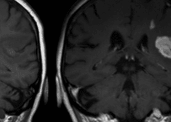
Impairments in gait and balance are not uncommon after stroke, and are associated with poorer functional recovery. The contralesional cerebellum is strongly implicated in the functional reorganization of the motor network when recovery takes place. In fact, magnetic resonance imaging (MRI) studies have shown that activity in the contralesional cerebellum is positively associated with gait recovery in patients with stroke. Cerebellar mediated motor learning can be potentiated using non-invasive brain stimulation methods, including cerebellar intermittent θ-burst stimulation (CRB-iTBS). In this randomized controlled trial, 34 patients were randomized to receive a 3-week treatment of CRB-iTBS coupled with physiotherapy or sham iTBS, to determine whether CRB-iTBS can improve balance and gait functions in patients with hemiparesis due to stroke. The primary outcome was the between-group difference in change from baseline in the Berg Balance Scale. Secondary exploratory measures included the between-group difference in change from baseline in Fugl-Meyer Assessment scale, Barthel Index, and locomotion assessment with gait analysis and cortical activity measured by transcranial magnetic stimulation in combination with electroencephalogram. Researchers found that patients treated with CRB-iTBS, but not with sham iTBS, showed an improvement in gait and balance functions, as revealed by a pronounced increase in the mean Berg Balance Scale score (p<0.001). No overall treatment-associated differences were noted in the Fugl-Meyer Assessment. In addition, patients treated with CRB-iTBS, but not sham iTBS, showed a reduction in step width from a baseline mean of 16.8 cm to 14.3 cm after 3 weeks of treatment (p<0.05). Increased neural activity over the posterior parietal cortex was also noted in the intervention group. This study therefore shows that cerebellar intermittent θ-burst stimulation may promote gait and balance recovery in patients affected by stroke.
Image: PD
©2018 2 Minute Medicine, Inc. All rights reserved. No works may be reproduced without expressed written consent from 2 Minute Medicine, Inc. Inquire about licensing here. No article should be construed as medical advice and is not intended as such by the authors or by 2 Minute Medicine, Inc.


