2 Minute Medicine Rewind November 15, 2021
1. On chest x-ray, a cardiothoracic ratio (CTR) of greater than 0.5 is an indeterminate indicator for cardiac enlargement.
2. Optimal cut-off points for identifying cardiac enlargement on x-ray were calculated, taking into account sex, heart diameter, and CTR.
Evidence Rating Level: 2 (Good)
Cardiac enlargement is an important radiological indicator of cardiac disease. Although cardiac magnetic resonance imaging (MRI) is the most accurate evaluator of cardiac size, chest X-rays are used as first-line imaging for suspected cardiac or pulmonary conditions, due to its low cost and availability. On chest x-ray, the traditional method of identifying cardiac enlargement is with a cardiothoracic ratio (CTR) of greater than 0.5: However, some studies demonstrated low diagnostic accuracy with this cut-off, and this method has not been validated against cardiac MRI. Therefore, this current retrospective study aimed to identify the validity of various cut-offs for cardiac enlargement on chest x-ray, using heart diameter cut-offs and CTR cut-offs. The study population consisted of 152 patients, with a mean (SD) age of 52 (17), who had a chest radiograph followed by a cardiac MRI within 14 days. The results showed that using the 0.5 CTR cut-off point yielded a 72% sensitivity and 72% specificity. However, with posteroanterior (PA) radiographs, the optimal cut-offs for ruling out cardiac enlargement were a CTR of 0.45 (88% sensitivity, 0.31 negative likelihood ratio), a heart diameter of 15 cm for men (85% sensitivity, 0.24 negative LR), and diameter of 13 cm for women (91% sensitivity, 0.15 negative LR). For ruling in cardiac enlargement, the optimal cut-offs were 0.60 CTR (94% specificity, 7.80 positive LR), 19 cm diameter in men (100% specificity, positive LR of infinity), and 17 cm in women (91% specificity, positive LR of 3.5). Although anteroposterior (AP) radiographs had significantly higher CTRs and diameters, the optimal cut-offs were found to be the same. Overall, this has shown that the traditional CTR cut-off of 0.5 is indeterminate for identifying cardiac enlargement, and taking into account sex, heart diameter, and CTR to develop cut-offs will enhance diagnostic accuracy.
1. For in-hospital cardiac arrest with shockable rhythms, epinephrine use before first defibrillation was associated with lower rates of survival at discharge, lower rates of neurological survival at discharge, and survival after acute resuscitation, independent of the time taken to first defibrillation.
2. However, patients receiving early epinephrine were found to be less likely to be on mechanical ventilation or have a myocardial infarction during the same admission.
Evidence Rating Level: 2 (Good)
In the United States, guidelines for in-hospital cardiac arrest dictate that for patients with a shockable rhythm, defibrillation within two minutes is recommended, with epinephrine not used until after two defibrillation attempts. This guideline is due to the high effectiveness of restoring circulation with defibrillation, whereas the use of epinephrine is controversial, but currently recommended every three to five minutes for non-shockable rhythms. Despite these recommendations, a recent study found that 51% of patients with in-hospital cardiac arrest and shockable rhythms received epinephrine before the second defibrillation, which had a 30% reduction in survival odds. The current propensity matched analysis examined the prevalence of epinephrine use before first defibrillation, patient factors associated with higher frequency of epinephrine use, and outcomes such as survival to discharge, neurological function at discharge (using the cerebral performance category score), and survival after acute resuscitation (defined as restoration of circulation for 20 minutes). The data from this study was from the American Heart Association’s Get With The Guidelines-Resuscitation (GWTG-R) registry, a registry of in-hospital cardiac arrest. In total, the study population consisted of 34,820 patients with shockable rhythms, of which 27.7% received epinephrine before first defibrillation. This trend increased over time, with 20.8% of patients in 2000 increasing to 29.8% in 2018 (p for trend < 0.001). Patients who received epinephrine were less likely to be on mechanical ventilation or have a myocardial infarction during the same hospital admission, but were more likely to be black, have renal or respiratory insufficiency, have sepsis, or were on intravenous antiarrhythmic medications (p < 0.0001 for all). Additionally, patients receiving epinephrine had a lower survival to discharge rate (25.0% versus 18.4%, difference of 23.1%, 95% CI -24.1 to -22.0%), a lower rate of favourable neurological survival (18.4% versus 39.2%, difference of -20.8%, 95% CI -21.8 to -19.8%), and a lower rate of survival after acute resuscitation (64.2% versus 81.0%, difference of -16.9%, 95% CI -18.0 to -15.8%). In a sensitivity analysis, the worse outcomes persisted even after controlling for time taken to first defibrillation. Overall, this study demonstrated that epinephrine use prior to first defibrillation in shockable in-hospital cardiac arrest is associated with decreased survival and neurological function, independent of time taken to defibrillation.
1. Persistent physical symptoms during the COVID-19 pandemic was associated with believing to have been infected with SARS-CoV-2, and not associated with laboratory-confirmed infection, apart from persistent anosmia.
Evidence Rating Level: 2 (Good)
Individuals infected with SARS-CoV-2 have a higher risk of persistent physical symptoms, colloquially referred to as “long COVID”. However, it is also possible that these symptoms may have its origin in other causes, as opposed to stemming from COVID-19 infection. This current cross-sectional analysis examined the association between persistent physical symptoms and self-reported belief of COVID-19 infection or serology test results. The hypothesis was that self-belief in having been infected would be associated with persistent symptoms, even when controlling for actual infection. The study population consisted of 26,823 individuals taken from the French CONSTANCES cohort study, consisting of volunteers aged 18 to 69 who responded to annual questionnaires. From May to November 2020, participants received self-sampling serology test kits, and were notified of their results. From December 2020 to January 2021, participants responded to a questionnaire about persistent physical symptoms, including pains and aches, sleep difficulties, sensory symptoms, breathing difficulties, digestive problems, anosmia, and headache. They were also asked if they believed they were infected previously and if this was confirmed by testing, and participants believing they were first infected after serologic testing were excluded. Overall, the prevalence of persistent symptoms ranged from .5% for anosmia to 10.2% for sleep difficulties. 914 participants believed they had a COVID-19 infection before the serology test, 49.6% of which had a positive serology test. After adjustment, a positive belief was associated with greater odds of all symptoms (except for hearing impairment and sleep difficulties) with odds ratios (ORs) ranging from 1.39 (95% CI 1.03-1.86) to 16.37 (95% CI 10.21-26.24). However, a positive serology test was only associated with greater odds for anosmia (OR 2.72, 95% CI 1.66-4.46) and was associated with lower odds for skin problems (OR 0.49, 95% CI 0.29-0.85). There was no significant interaction between belief and serology test, and similar results were present even after adjusting for self-rated health and depressive symptoms. In conclusion, this study showed how, apart from anosmia, persistent physical symptoms during the first months of the COVID-19 pandemic were associated with believing to have been infected, rather than having a lab confirmed infection.
1. For large-vessel occlusion stroke, there were no clinical or safety differences in outcomes for patients undergoing mechanical thrombectomy between 6 and 24 hours after symptom onset, when the imaging modality was non-contrast CT compared to more advanced imaging, such as CT perfusion and MRI.
Evidence Rating Level: 2 (Good)
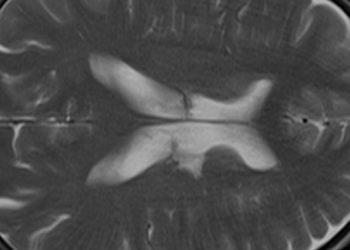
Due to the results of the DAWN and DEFUSE 3 trials, patients with large-vessel occlusion stroke can receive endovascular treatment if presenting 6-24 hours after symptom onset, which is an extended time period than was indicated previously. In these studies, magnetic resonance imaging (MRI) or computed tomography perfusion (CTP) were used as triage. However, these are advanced imaging modalities, and accessibility is limited. The purpose of this multinational cohort study was to determine if patients with proximal anterior circulation occlusion stroke, triaged with noncontrast CT (NCCT), would also benefit from endovascular mechanical thrombectomy (MT) in this extended time window, compared to those imaged with MRI or CTP. The hypothesis was that there would be no difference in outcomes between the two different imaging cohorts, and the main outcomes measured were scores at 90 days from the modified Rankin Scale (mRS) for Neurological Disability, 90-day functional independence (mRS scores of 0-2), symptomatic intracranial hemorrhage, and 90-day mortality. There were 1604 patients included in the study: 33.3% received NCCT imaging, 46.9% received CTP, and 19.8% received MRI. Individuals in the NCCT group had a higher median baseline National Institutes of Health Stroke Scale (NIHSS) score, higher rates of atrial fibrillation and hypertension, and were more likely to be transfers. The time from symptom onset to puncture was shorter in the NCCT group (median [IQR] of 10.4 [7.8-14.4] hours), compared to CTP (11.3 [8.4-15.2] hours) and MRI (12.4 [9.4-15.4] hours), with p < 0.001. As well, successful reperfusion rates were higher in the NCCT group (88.9%) compared to CTP and MRI (89.5% and 78.9% respectively, p < 0.001). For the 90-day mRS score, there was no difference between NCCT and CTP (adjusted odds ratio 0.95, 95% CI 0.77-1.17, p = 0.64) nor between NCCT and MRI (aOR 0.95, 95% CI 0.80-1.13, p = 0.55). There was also no difference for 90-day functional independence: the proportion of patients having an mRS score between 0 to 2 were 41.2% for NCCT, 44.3% for CTP, and 38.7% for MRI. Symptomatic intracranial hemorrhage was similar in all modalities (NCCT: 8.1%, CTP: 5.8%, MRI: 4.7%, p = 0.11) and 90-day mortality was also similar (NCCT: 23.4%, CTP: 21.1%, MRI: 19.5%, p = 0.38). Overall, this study demonstrated that there is no difference in clinical and safety outcomes for mechanical thrombectomy between 6 to 24 hours after symptom onset, when the imaging modality is with NCCT compared to CTP or MRI imaging.
1. A blended model of CBT (bCBT), consisting of face-to-face sessions alternated with online module sessions, was as clinically effective for treating anxiety disorders as solely face-to-face CBT (ftfCBT)
2. bCBT was equally as acceptable to patients and therapists, and was more cost-effective than ftfCBT for direct medical costs.
Evidence Rating Level: 2 (Good)
Cognitive behavioural therapy (CBT) is an effective treatment for anxiety disorders. However, fewer than 50% of people with anxiety disorders undergo appropriate treatments, with reasons including lack of therapists and high therapy costs. A proposed alternative to mitigate costs and improve accessibility is Internet-delivered CBT (iCBT), which consists of online modules with and without an online therapist assisting. However, there is reluctance for therapists to use iCBT due to low adherence, concerns about addressing complex symptoms, and not being able to form strong interpersonal relationships. Therefore, a blended CBT (bCBT) approach has been proposed, combining iCBT with face-to-face CBT (ftfCBT): A feasibility randomized controlled trial was done with 18 participants randomized to either treatment, finding no difference between these groups in reduction of anxiety symptoms. The current randomized controlled trial aimed to investigate the acceptability, cost-effectiveness, and clinical effectiveness of bCBT compared to ftfCBT, for patients with panic disorder (PD), social anxiety disorder (SAD), and generalized anxiety disorder (GAD) in outpatient care. In total, there were 52 patients randomized to bCBT and 62 to ftfCBT, with a mean (SD) age of 36.3 (10.6). The bCBT group received 15 weekly sessions alternating between face-to-face and online modules with asynchronous feedback from the therapist. Questionnaires at baseline, week 7 (mid-treatment), week 15 (post-treatment), and 1 year follow-up were used to assess the outcomes of acceptability (such as treatment adherence, treatment preference, and satisfaction) and clinical effectiveness (using the Beck Anxiety Inventory (BAI)). The results showed that adherence was not significantly different between the groups, with bCBT being slightly higher (67.4% versus 61.6% completing all sessions, p = 0.608). There was also no difference in ratings of the therapeutic alliance, using the Working Alliance Inventory (WAI-SR), from both the patients’ (p = 0.912) and therapists’ perspectives (p = 0.762). Treatment satisfaction was also not different (p = 0.750), with the mean Client Satisfaction Questionnaire (CSQ-8) scores for both falling between “somewhat satisfied” and “very satisfied”. Furthermore, there were no significant differences in BAI score, representing decreased anxiety severity, between the two groups post-treatment and at 1-year follow-up (p = 0.477 and 0.093 respectively). In terms of cost-effectiveness, bCBT was significantly cheaper for direct medical costs, which were €3758 for bCBT and €3841 for ftfCBT during the 1-year period (mean difference -83.78, 95% CI -96.96 to -70.61, p < 0.001). For societal costs, which included medical, patient, and productivity costs, were not different between the two treatments (€10,945 for bCBT, €10,937 for ftfCBT, mean difference 26.46, 95% CI -24.64 to 42.71, p > 0.01). Overall, this study demonstrated that bCBT is as clinically effective as ftfCBT for anxiety disorders, and is equally acceptable to patients and therapists while being more cost-effective in terms of direct medical costs. However, further randomized trials with greater sample sizes will be needed to strengthen and validate these findings.
Image: PD
©2021 2 Minute Medicine, Inc. All rights reserved. No works may be reproduced without expressed written consent from 2 Minute Medicine, Inc. Inquire about licensing here. No article should be construed as medical advice and is not intended as such by the authors or by 2 Minute Medicine, Inc.


