


Computer modeling improves orbital fracture repair
Jan 15th – CT-generated mirror image overlay (MIO) technique results in significant reduction in postoperative diplopia, reoperation.
![]() [tabs tab1=”2MM Rundown” tab2= “2MM Full Report” tab3=”About the Authors”]
[tabs tab1=”2MM Rundown” tab2= “2MM Full Report” tab3=”About the Authors”]
[tab]
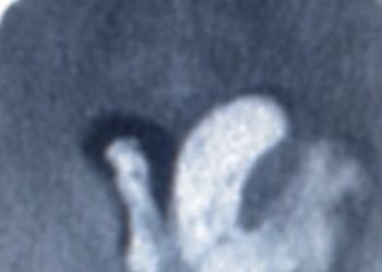
Image: CC/J.Heilman
1. The CT-generated mirror image overlay (MIO) technique results in significant reduction in postoperative diplopia in patients with complex orbital fractures.
2. MIO significantly reduces the incidence of abnormal globe positioning and enophthalmos postoperatively.
3. The need for revision surgery is reduced from 20% to 4% by implementing the MIO protocol.
This study demonstrated that MIO has many benefits when used for the repair of unilateral orbital fractures. The incidence of diplopia, enophthalmos, and need for repeat surgery were all significantly decreased in patients who received MIO treatment. Limitations to this study include the non-differentiation between diplopia based on orbital position and that due to neuromuscular etiologies. The retrospective nature of the study, in addition to the fact that only one physician evaluated measures of proper globe position, introduced a setting in which a large degree of bias may be possible. Finally, the measurement of diplopia was performed using a subjective, reporting depended scale. A technique such as perimetry would have been more objective and sensitive.
Click to read the study in JAMA Facial Plastic Surgery
[/tab]
[tab]
Image: CC/J.Heilman
1. The CT-generated mirror image overlay (MIO) technique results in significant reduction in postoperative diplopia in patients with complex orbital fractures.
2. MIO significantly reduces the incidence of abnormal globe positioning and enophthalmos postoperatively.
3. The need for revision surgery is reduced from 20% to 4% by implementing the MIO protocol.
Primer: Orbital fractures are not often life threatening; however, they can carry significant morbidity with regards to vision and cosmesis. Visual deficits such as diplopia and blindness can be induced by a range of traumatic etiologies including globe rupture, extraocular muscle entrapment, or direct CNS injury. Cosmetic appearance also suffers as a result of injury and bony deformation. Urgent intervention is often required in order to assure that function and aesthetics are maintained. Reconstruction of orbital fractures can be technically challenging. The most common injury is blowout of the infraorbital floor due to elevated intraorbital pressures. These types of injuries necessitate access to a confined space where lighting, field of vision, and working space is limited. Additionally, injuries in the areas of the zygomatic-maxillary and nasal-ethmoid regions cause significant disruption to the orbit which requires complex reconstruction with fixation equipment to regain prior facial structure.
It has been shown that the most important aspect of reducing postoperative morbidity is the proper repositioning of the orbital bones. The MIO technique requires that a patient receive a CT scan before operation in order to discern the bony structure prior to the traumatic event. This is done by utilizing a computer-generated mirror image of the non-traumatized half of the skull as a guide during operative reconstruction. The CT based guidance information can be loaded onto a computer in the operating room and an adhesive mask can be applied to the patient in order to sync the navigational model to 3-D space. After this is accomplished a probe can be utilized to ascertain proper repositioning of deformities and correct locations for implantable supports.
Background reading:
1. Orbital fractures [UptoDate]
This [retrospective cohort] study: utilized 90 patients from a consecutive cohort who had suffered unilateral traumatic injury to the orbit. The 45 members of the control group were gathered prior to the MIO group and received traditional non-MIO guided reconstruction. All patients received reconstruction from or directly supervised by the same surgeon. The results of this study show that patients in the MIO group had a 2-fold improvement in postoperative diplopia over controls, a significant improvement (p=.003). Abnormal globe positions, as assessed by the reconstructive surgeon, were found to be significantly improved in 88% of MIO patients as opposed to 69% of controls. Finally, the need for repeat surgery was shown to be decreased from 20% to 4% with the use of MIO compared to control operation (p=.03).
In sum: This study demonstrated that MIO has many benefits when used for the repair of unilateral orbital fractures. The incidence of diplopia, enophthalmos, and need for repeat surgery were all significantly decreased in patients who received MIO treatment. Limitations to this study include the non-differentiation between diplopia based on orbital position and that due to neuromuscular etiologies. The retrospective nature of the study, in addition to the fact that only one physician evaluated measures of proper globe position, introduces a setting in which a large degree of bias may be possible. Finally, the measurement of diplopia was performed using a subjective, reporting depended scale. A technique such as perimetry would have been more objective and sensitive.
Click to read the study in JAMA Facial Plastic Surgery
By [DM] and [MK]
© 2013 2minutemedicine.com. All rights reserved. No works may be reproduced without written consent from 2minutemedicine.com. Disclaimer: We present factual information directly from peer reviewed medical journals. No post should be construed as medical advice and is not intended as such by the authors or by 2minutemedicine.com. PLEASE SEE A HEALTHCARE PROVIDER IN YOUR AREA IF YOU SEEK MEDICAL ADVICE OF ANY SORT. Content is produced in accordance with fair use copyrights solely and strictly for the purpose of teaching, news and criticism. No benefit, monetary or otherwise, is realized by any participants or the owner of this domain.
[/tab]
[tab]
 Devin Miller: Devin is a 3rd year M.D. candidate at Johns Hopkins University.
 Mimmie Kwong: Mimmie is a 3rd year M.D. candidate at Johns Hopkins School of Medicine in Baltimore, MD.
[/tab]
[/tabs]
RelatedReports
