Infrared thermography reliably measures growth of infantile hemangiomas
1. Infrared thermography of 42 infantile hemangiomas indicated that the lesions peak in temperature at 3 months and progressively decrease thereafter, a change that was correlated with a subjective visual analog scale score.
2. Infrared thermography reliably measured the temperature of a cutaneous infantile hemangioma, showing no significant difference between a baseline measurement and a measurement taken after 30 minutes.
Evidence Rating Level: 2 (Good)
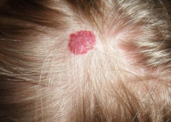
Study Rundown: Infantile hemangiomas (IHs) are common cutaneous vascular tumors of infancy that often occur on the scalp, face, and neck. Although the tumors are benign, they have the potential to cause significant disfigurement. An IH will go through phases of proliferation and eventual involution. Effective management of the IH requires understanding where it lies on this natural course. This is typically determined by subjective assessment of its color, size, tenseness, and temperature reported on a visual analog scale (VAS), although this measure is not validated. The authors investigated the role of infrared thermography (IRT), which can objectively measure local temperature differences, in assessing IHs. An examination of 42 infants revealed that IRT was a reliable measure of IH temperature, and temperature differences were well correlated with the subjectively determined VAS score (r = -0.25), indicating that infrared thermography was a reliable and objective method for assessing IHs well suited for daily clinical practice. The study was limited by its small sample size.
Click to read the study in JAMA Dermatology
Relevant Reading: Growth characteristics of infantile hemangiomas
In-Depth [prospective cohort]: This study evaluated 42 infants (including 35 females) with a mean age of 3.7 months. Infants had IHs on the head and neck, trunk, extremities, and genital area. At each study visit, the local temperature of each IH was measured using a digital IRT device by touching the probe to the surface of the lesion for a few seconds and then again after 30 minutes (as a test for reliability). The skin temperature of the contralateral unaffected side was measured for comparison. A blinded assessor independently reviewed photographs of the IHs to provide a VAS score for each lesion at every visit. The temperature difference (temperature of the IH minus
temperature of the contralateral skin) for each patient was compared with the changes in the VAS over time. The mean temperature difference at baseline was 1.9°F, and peaked to 2.5°F at 3 months. Change in temperature as measured by the IRT was inversely correlated with mean visual analog scale (r = −0.25), indicating a strong correlation between the IRT’s temperature assessment and a subjectively determined VAS score. Mean temperature differences recorded at baseline and 30 minutes later were not significant (p=0.14), suggesting good reliability of IRT.
More from this author: Systematic reviews moderately reflect disease burden in dermatology, Bath psoralen plus ultraviolet A shows good efficacy in the treatment of mycosis fungoides, Lower rates of self skin examination among ethnic minorities
Image: PD
©2012-2014 2minutemedicine.com. All rights reserved. No works may be reproduced without expressed written consent from 2minutemedicine.com. Disclaimer: We present factual information directly from peer reviewed medical journals. No post should be construed as medical advice and is not intended as such by the authors, editors, staff or by 2minutemedicine.com. PLEASE SEE A HEALTHCARE PROVIDER IN YOUR AREA IF YOU SEEK MEDICAL ADVICE OF ANY SORT.


![Childhood ADHD associated with increased risk of suicide [Physician Comment]](https://www.2minutemedicine.com/wp-content/uploads/2013/03/PET-image1-e1377449984183-75x75.jpg)

