
MRI study characterizes breast cancer patterns in augmented breasts
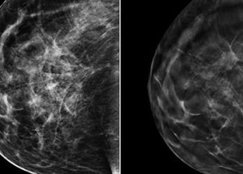
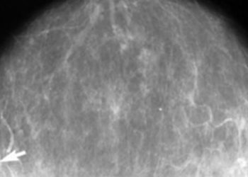
1. Magnetic resonance imaging examination of patients with proven breast carcinoma in silicone or saline augmented breasts visualized tumor spread along the implant contour and abutting the pectoralis major, most commonly in retroglandular implants versus retropectoral implants. However, neither implant type was associated with a difference in tumor invasion of the pectoralis.
2. Magnetic resonance imaging was able to reveal a greater extent of disease than mammography or ultrasound in nearly half of the cases, and detected roughly a third of the index carcinomas plus a small number of unknown contralateral carcinomas.
Evidence Rating Level: 2 (Good)
Study Rundown: Elective breast augmentation continues to be an increasingly popular surgical procedure, and as the number of women with breast implants increases, proper screening and characterization to recognize breast carcinomas in these women is of greater importance. Although mammography and ultrasonography are the standard of care in breast cancer screening, magnetic resonance imaging (MRI) is a useful adjunct imaging modality for the characterization of breast anatomy. Due to its high spatial resolution and ability to better examine soft tissues, MRI is potentially well-suited to the evaluation of augmented breasts due to the compression of normal breast tissue by the implant, and may even be valuable in the presurgical breast assessment. The present study sought to characterize the MRI features of breast carcinomas detected in women with breast implants through an MRI database review of patients with biopsy-confirmed breast cancer in augmented breasts. The authors found that breast tumors often contacted either the implant or the pectoralis major muscle, with growth abutting the implant contour more frequently in breasts with retroglandular implants than those with retropectoral implants. While MRI may be a valuable adjunct to mammography and ultrasound in characterizing and evaluating disease, it does not detect nodal disease as well as other modalities, but it was able to reveal a greater extent of tumor than mammography or ultrasound in 40% of cases. However, the appropriateness of MRI as a primary screening technique remains tenuous as it was only able to detect 30% of the index carcinomas. The primary limitations of this study were its small sample size, and retrospective design which may have introduced a degree of selection bias. Further investigations are warranted into the determining the most appropriate multimodality detection technique and prognosis of breast cancers in patients with implants.
Click to read the study in American Journal of Roentgenology
Relevant Reading: Breast cancer diagnosis in women with subglandular silicone gel-filled augmentation implants.
In-Depth [retrospective cohort]: A total of 54 consecutive patients with both biopsy-confirmed breast carcinoma and ipsilateral breast augmentation were identified from a database of patients who had undergone breast MRI between 2000 and 2010. Patients included in the study had a median age of diagnosis at 49 years (range: 27—72 years) and had their breast implants in place for an average 10 years before receiving a breast cancer diagnosis. Images from the identified patients were reviewed for type and location of the implant and tumor characteristics were identified. Of the 54 cancers, the mean tumor size was 2.8 cm (range: 0.6–9.6 cm) and the location of the implants was retropectoral in 38 (70%) and retroglandular in 16 (30%) and 38 of the 54 (70%) cancers were palpable. The cases included 46 (85%) invasive cancers (invasive ductal carcinoma, n = 35; invasive lobular carcinoma, n = 7; and mixed features, n = 4) and eight (15%) ductal carcinomas in situ. No significant difference was found between implant position and lesion morphology (p = 0.55) or tumor size (p = 1.00). Twenty of 54 (37%) carcinomas spread alongside the implant, 13 (24%) bordered the pectoralis major muscle, and 2 (4%) invaded the pectoralis major muscle. Of the tumors bordering the implant, 90% spread along the implant capsule and this pattern was more common in women who had received retroglandular implants (56%) than retropectoral implants (24%) (p = 0.03). Across all patients, MRI was only able to detect the index carcinoma in 16 (30%) patients, though it was able to reveal a greater extent of disease than that seen on ultrasound or mammography in 21 of 52 patients (40%). Additionally, MRI revealed previously unsuspected carcinoma in the contralateral breast in 3 of 54 cases (6%).
Image: PD
©2015 2 Minute Medicine, Inc. All rights reserved. No works may be reproduced without expressed written consent from 2 Minute Medicine, Inc. Inquire about licensing here. No article should be construed as medical advice and is not intended as such by the authors or by 2 Minute Medicine, Inc.


