
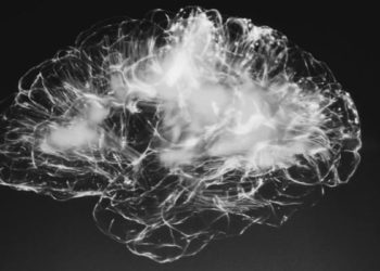
PET imaging of tau may help distinguish Alzheimer Disease from other neurodegenerative disorders
1. In this cross-sectional study, the tau marker [18F]flortaucipir accurately distinguished Alzheimer’s Disease (AD) dementia from other non-AD neurodegenerative disorders about 90% of the time.
2. [18F]flortaucipir imaging more accurately identified AD compared to MRI-based measures of brain atrophy specific associated with AD.
Evidence Rating Level: 2 (Good)
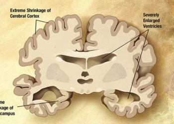
Study Rundown: Alzheimer’s Disease (AD) is typified by both amyloid-beta and tau pathology, and new imaging techniques with radiotracers to these molecules have been suggested as biomarkers of the disease. Though amyloid-beta based imaging techniques were developed first, amyloid pathology precedes cognitive decline, and many older patients show significant amyloid burden despite being cognitively intact, impairing the usefulness of this imaging technique for diagnosing AD. In this cross-sectional study, a new PET imaging-based tau marker, [18F]flortaucipir, showed about a 90% accuracy rate of distinguishing AD from other neurodegenerative disorders. Though not as robust, [18F]flortaucipir imaging was able to identify cases of MCI due to AD pathology, which is considered a prodromal stage of AD. Further, [18F]flortaucipir imaging more accurately identified AD compared to MRI-based measures of brain atrophy associated with AD.
Though this study suggests [18F]flortaucipir may be a useful clinical tool in the future, the study’s results are difficult to interpret without post-mortem validation of AD cases. In addition, patients were recruited from memory disorder clinics and already had established diagnoses, representing a potential source of bias. Finally, some of the neurodegenerative disorders used for comparison did not have a dementia component as a control, and it is unclear what the accuracy of this imaging test in more “real-world” situations where the definitive cause of the dementia is in doubt.
Click to read the study in JAMA
Relevant Reading: Tau PET patterns mirror clinical and neuroanatomical variability in Alzheimer’s disease
In-Depth [cross-sectional study]: This multicenter, cross-sectional study recruited patients with a neurodegenerative disorder (179 with AD, 254 with non-AD neurodegenerative disorders, 83 with MCI due to AD, 43 non-AD MCI, and 160 cognitively normal controls) from memory disorder clinics in Seoul, Sweden, and San Francisco between June 2014 and November 2017. AD dementia was defined as typical symptoms with verification of amyloid-beta in the CSF or with amyloid-beta PET imaging. The temporal meta-ROI for [18F]flortaucipir imaging distinguished AD from controls with 90.3% (CI95 87.1 to 92.9%) accuracy, 89.9% (CI95 84.6 to 93.9%) sensitivity, and 90.6% (CI95 86.3 to 93.9%) specificity. The temporal meta-ROI for [18F]flortaucipir imaging distinguished AD MCI from controls with 83.4% (CI95 79.0 to 87.2%) accuracy, 61.5% (CI95 50.1 to 71.9%) sensitivity, and 90.6% (CI95 86.3 to 93.9%) specificity. [18F]flortaucipir imaging more accurately predicted AD compared to MRI-based measures of brain atrophy specific for AD (p < 0.001).
Image: PD
©2018 2 Minute Medicine, Inc. All rights reserved. No works may be reproduced without expressed written consent from 2 Minute Medicine, Inc. Inquire about licensing here. No article should be construed as medical advice and is not intended as such by the authors or by 2 Minute Medicine, Inc.


![siRNA against antithrombin alleviates symptoms of hemophilia [PreClinical]](https://www.2minutemedicine.com/wp-content/uploads/2015/04/clot-CCWiki-75x75.jpg)