
Pulmonary MRI with ultrashort echo time is comparable to pulmonary CT
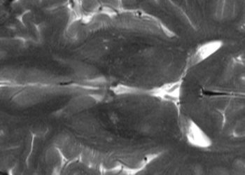
1. Pulmonary magnetic resonance imaging (MRI) with ultrashort echo time (UTE) is a noninvasive method of evaluation of the lung parenchyma, providing regional morphologic and functional assessments of various pulmonary diseases in a manner comparable to conventional computed tomography (CT) without radiation exposure.
2. MRI with UTE provides substantially improved pulmonary parenchymal enhancement for the characterization of non-uniform disruption of lung architecture such as that seen in emphysema, bronchiectasis, and reticular opacifications as compared to conventional pulmonary CT imaging.
Evidence Rating Level: 2 (Good)
Study Rundown: Pulmonary diseases remain a devastating cause of morbidity and mortality worldwide, with limited treatment options for many conditions. Management of lung disease is challenging in part due to the limitations intrinsic to established diagnostic methods, such as radiation exposure in CT. Unlike CT, MRI does not involve exposure to ionizing radiation to provide high-resolution morphological and functional information; thus, it holds potential to provide safe and comprehensive lung disease evaluation. However, achieving the desired signal intensity on MRI is challenging due to characteristics intrinsic to lung tissue such as low proton density and multiple air-tissue interfaces, in addition to rapid signal decay causing traditional MR sequences to very poorly evaluate the lung parenchyma. The application of UTE sequences to pulmonary MRI has been suggested as a strategy to achieve a higher signal-to-noise ratio by dampening the effect of signal decay during image acquisition. This study evaluated the diagnostic accuracy of pulmonary MRI in the evaluation of lung diseases obtained using UTE MRI and compared the findings with standard CT assessments. The lung parenchyma and mediastinum were scanned using MRI and CT in patients presenting with various lung diseases and visually evaluated for detection and characterization of non-uniform disruption of tissue architecture using a 5-point system. This study was limited by its small cohort size and heterogeneity within the sample with respect to pulmonary disease features and patient demographic data, however it did find highly significant intermethod agreement in findings between the two imaging modalities. Evaluation of emphysematous changes, bullae, reticular opacities and bronchiectasis was superior with UTE MRI as compared to CT imaging. Additional studies focused on MRI evaluations of specific lung diseases would strengthen these findings in a larger patient population.
Click to read the study in the Journal of Magnetic Resonance Imaging
Relevant Reading: Lung morphology assessment with balanced steady-state free precession MR imaging compared with CT
In-Depth [prospective cohort]: Standard and low-dose CTs and MRI with UTE of the lungs and mediastinum were performed on 85 patients who presented with various lung diseases. Visual assessments of the images obtained followed a 5-point system to note presence of nodules or masses in addition to noting irregular tissue features such as ground-glass and reticular opacity, consolidation, bullae, bronchiectasis, reticular opacity, and honeycombing. The 5-point system was also used to account for pathological features within the mediastinum such as aneurysms, pleural/pericardial effusions, pleural thickening or tumor, and lymphadenopathy. When the diagnostic performance of the imaging methods were compared using kappa statistics and χ2 test, significant intermethod agreement was found (0.67 ≤ κ ≤ 0.98; P < 0.0001). Diagnostic accuracy of bullae suggestive of emphysema, bronchiectasis, and reticular opacity using standard-dose CT was stronger than assessments generated using low-dose CT (emphysema or bullae: p = 0.0002; reticular opacity: p < 0.0001). These features were even more prominent on pulmonary MRI (emphysema or bullae: p < 0.0001; bronchiectasis: p = 0.008; reticular opacity: p < 0.0001).
Image: CC/WikimediaCommons/Patrick J. Lynch
©2015 2 Minute Medicine, Inc. All rights reserved. No works may be reproduced without expressed written consent from 2 Minute Medicine, Inc. Inquire about licensing here. No article should be construed as medical advice and is not intended as such by the authors or by 2 Minute Medicine, In
RelatedReports


