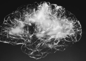
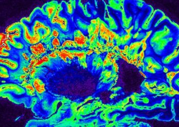
Regional central nervous damage associated with fatigue in multiple sclerosis
1. Multiple sclerosis (MS) patients with fatigue symptoms showed marked regional changes on magnetic resonance imaging (MRI) as compared to both non-fatigue MS patients and matched controls.
2. MRI failed to demonstrate a correlation between global brain atrophy and fatigue symptoms, suggesting that damage to specific brain structures is likely contributory.
Evidence Rating Level: 2 (Good)
Study Rundown: Multiple sclerosis (MS) is an autoimmune demyelinating disorder that causes progressive CNS damage with substantial resultant morbidity. Fatigue is one of the most common symptoms of MS and can significantly limit a patient’s ability to perform activities of daily living. Despite this, the underlying cause of fatigue among MS patients is poorly understood, and objective imaging assessments that can be used to broadly analyze and characterize fatigue are needed.
In this study, MRI was used to evaluate the brains of MS patients with and without fatigue. When compared to control patients, MS patients showed significant global brain atrophy, but no global atrophy differences were found between MS patients with respect to fatigue. However, imaging measures of regional brain atrophy showed distinguishing changes between the fatigued and non-fatigued MS groups, suggesting that fatigue is a function of damage to specific brain regions. The study’s primary limitation was its cross-sectional nature and lack of functional MR imaging findings that may have defined any pathogenetic link between presence of atrophy and fatigue severity.
Click to read the study in Radiology
Relevant Reading: Contribution of global and regional damage of the gray and white matter to fatigue in MS
In-Depth [cross-sectional study]: In this study, MRI was obtained in 63 consecutive patients with MS (31 fatigued, 32 non-fatigued) and 35 age- and sex-matched control subjects. All enrolled patients underwent neurological evaluations, including the Fatigue Severity Scale (FSS). Standard anatomic MRI was used alongside diffusion tensor imaging (DTI) to assess for damage and atrophy in grey matter (GM) and white matter (WM), both globally and in specific subcortical nuclei. As compared to controls, MS patients showed significantly greater GM and WM atrophy globally; no differences were found between fatigued and non-fatigued MS patients. Measurements of regional brain atrophy were significantly correlated with FSS scores (P < .001), specifically in the right nucleus accumbens, right inferior temporal gyrus (RITG), and left superior frontal gyrus. Using DTI, fatigued patients also showed a significantly greater degree of damage within the forceps major, left inferior frontooccipital fasciculus, and right anterior thalamic radiation (RATR). Using multivariate modeling, RITG atrophy and RATR damage were shown to be independent predictors of patient fatigue (P = .009 and P = .003, respectively).
Image: CC/Wiki
©2012-2014 2minutemedicine.com. All rights reserved. No works may be reproduced without expressed written consent from 2minutemedicine.com. Disclaimer: We present factual information directly from peer reviewed medical journals. No post should be construed as medical advice and is not intended as such by the authors, editors, staff or by 2minutemedicine.com. PLEASE SEE A HEALTHCARE PROVIDER IN YOUR AREA IF YOU SEEK MEDICAL ADVICE OF ANY SORT.


